|
Lesson 10.03 – Human Inheritance
Standards: B2.a, B2.b, B2.c, B2.d, B2.e, B2.f, B2.g, B3.a, B3.b, B7.b
INTRODUCTION
Of all the living things that inhabit this remarkable world, there is one in particular that has always drawn our interest, one that has always made us wonder, one that will always fire our imagination. That creature is, of course, ourselves, Homo sapiens.
Scientists once knew much less about humans than about other organisms. Until very recently, human genetics lagged far behind the genetics of “model” organisms such as fruit flies and mice. That, however, has changed. Scientists are now on the verge of understanding human genetics at least as well as they understand that of some other organisms. From that understanding will come a new responsibility to use that information wisely.
INSTRUCTION
Human Chromosomes
What makes us human? Biologists can begin to answer that question by taking a look under the microscope to see what is inside a human cell. To analyze chromosomes, cell biologists photograph cells in mitosis, when the chromosomes are fully condensed and easy to see. The biologists then cut out the chromosomes from the photographs and group them together in pairs. A picture of chromosomes arranged in this way is known as a karyotype (KAR-ee-uh-typ).
The chromosomes shown below are from a typical human body cell. The number of chromosomes—46—helps identify this karyotype as human. This karyotype is the result of a haploid sperm, carrying just 23 chromosomes, fertilizing a haploid egg, also with 23 chromosomes. The diploid zygote, or fertilized egg, contained the full complement of 46 chromosomes.

Karyotype These human chromosomes have been cut out of a photograph and arranged to form a karyotype.
Two of those 46 chromosomes are known as sex chromosomes, because they determine an individual's sex. Females have two copies of a large X chromosome. Males have one X and one small Y chromosome. To distinguish them from the sex chromosomes, the remaining 44 chromosomes are known as autosomal chromosomes, or autosomes. To quickly summarize the total number of chromosomes present in a human cell, both autosomes and sex chromosomes, biologists write 46,XX for females and 46,XY for males.
Click here (http://www.youtube.com/watch?v=8YMhkweABfQ&feature=related) to watch a video of how sex is determined in humans.
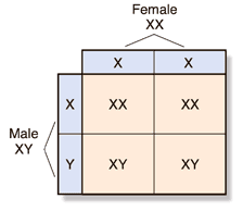
As you can see in the figure below, males and females are born in a roughly 50 : 50 ratio because of the way in which sex chromosomes segregate during meiosis. All human egg cells carry a single X chromosome (23,X). However, half of all sperm cells carry an X chromosome (23,X) and half carry a Y chromosome (23,Y). This ensures that just about half of the zygotes will be 46,XX and half will be 46,XY.
Segregation of Sex Chromosomes
In humans, egg cells contain a single X chromosome. Sperm cells contain either one X chromosome or one Y chromosome. In a population, approximately half of the zygotes are XX (female) and half are XY (male).
Genes and the Environment
Unfortunately for folks who would like to settle burning issues, like which side of the family is responsible for your good looks, some of the most obvious human traits are almost impossible to associate with single genes. There are two reasons for this. First, things you might think of as single traits, such as the shape of your eyes or ears, are actually polygenic, meaning they are controlled by many genes. Second, many of your personal traits are only partly governed by genetics. Remember that the phenotype of an organism is only partly determined by its genotype. Many traits are strongly influenced by environmental, or nongenetic, factors, including nutrition and exercise. For example, even though a person's maximum possible height is largely determined by genetic factors, nutritional improvements in the United States and Europe have increased the average height of these populations about 10 centimeters over their average height in the 1800s.
Although it is important to consider the influence of the environment on the expression of some genes, it must be understood that environmental effects on gene expression are not inherited; genes are. Genes may be denied a proper environment in which to reach full expression in one generation. However, these same genes can, in a proper environment, achieve full potential in a later generation.
Blood Group Genes
Human blood comes in a variety of genetically determined blood groups. Knowing a person's blood group is critical because using the wrong type of blood for a transfusion during a medical procedure can be fatal. A number of genes are responsible for human blood groups, but the best known are the ABO blood groups and the Rh blood groups.
The Rh blood group is determined by a single gene with two alleles—positive and negative. Rh stands for “rhesus monkey,” the animal in which this factor was discovered. The positive (Rh+) allele is dominant, so persons who are Rh+/Rh+ or Rh+/Rh− are said to be Rh-positive. Individuals with two Rh− alleles are Rh-negative.
The ABO blood group is more complicated. There are three alleles for this gene, IA, IB, and i. Alleles IA and IB are codominant. These alleles produce molecules known as antigens on the surface of red blood cells. As the table below shows, individuals with alleles IA and IB produce both A and B antigens, making them blood type AB. The i allele is recessive. Individuals with alleles IAIA or IAi produce only the A antigen, making them blood type A. Those with IBIB or IBi alleles are type B. Those who are homozygous for the i allele (ii) produce no antigen, and are said to have blood type O.
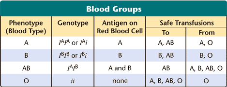
Blood Groups This table shows the relationship between genotype and phenotype for the ABO blood group. It also shows which blood types can safely be transfused into people with other blood types.
Recessive Alleles
Many human genes have become known through the study of genetic disorders. The table at right lists some common genetic disorders. In most cases, the presence of a normal, functioning gene is revealed only when an abnormal or nonfunctioning allele affects the phenotype.
One of the first genetic disorders to be understood this way was phenylketonuria (fen-ul-ket-oh-NOOR-ee-uh), or PKU. People with PKU lack the enzyme that is needed to break down phenylalanine. Phenylalanine is an amino acid found in milk and many other foods. If a newborn has PKU, phenylalanine may build up in the tissues during the child's first years of life and cause severe mental retardation. Fortunately, newborns can be tested for PKU and then placed on a low-phenylalanine diet that prevents most of the effects of PKU. PKU is caused by an autosomal recessive allele carried on chromosome 12.
Many other disorders are also caused by autosomal recessive alleles. One is Tay-Sachs disease, which is caused by an allele found mostly in Jewish families of central and eastern European ancestry. Tay-Sachs disease results in nervous system breakdown and death in the first few years of life. Although there is no treatment for Tay-Sachs disease, there is a test for the allele. By taking this test, prospective parents can learn whether they are at risk of having a child with the disorder.
Dominant Alleles
Not all genetic disorders are caused by recessive alleles. You may recall that the effects of a dominant allele are expressed even when the recessive allele is present. Therefore, if you have a dominant allele for a genetic disorder, it will be expressed. Two examples of genetic disorders caused by autosomal dominant alleles are a form of dwarfism known as achondroplasia (ay-kahn-droh-PLAY-zhuh) and a nervous system disorder known as Huntington disease. Huntington disease causes a progressive loss of muscle control and mental function until death occurs. People who have this disease generally show no symptoms until they are in their thirties or older, when the gradual damage to the nervous system begins.
Codominant Alleles
Sickle cell disease, a serious disorder found in about 1 out of 500 African Americans, is caused by a codominant allele. As you will read, the reason for the high incidence of sickle cell in the United States is a story that links genetics, human history, and molecular biology.
Sex-Linked Genes
Is there a special pattern of inheritance for genes located on the X chromosome or the Y chromosome? The answer is yes. Because these chromosomes determine sex, genes located on them are said to be sex-linked genes. Many sex-linked genes are found on the X chromosome, as shown in the figure at right. More than 100 sex-linked genetic disorders have now been mapped to the X chromosome. The human Y chromosome is much smaller than the X chromosome and appears to contain only a few genes.
Colorblindness
Three human genes associated with color vision are located on the X chromosome. In males, a defective version of any one of these genes produces colorblindness, an inability to distinguish certain colors. The most common form of this disorder, red-green colorblindness, is found in about 1 in 10 males in the United States. Among females, however, colorblindness is rare—only about 1 female in 100 has colorblindness. Why the difference?
Males have just one X chromosome. Thus, all X-linked alleles are expressed in males, even if they are recessive. In order for a recessive allele, such as the one for colorblindness, to be expressed in females, there must be two copies of the allele, one on each of the two X chromosomes. This means that the recessive phenotype of a sex-linked genetic disorder tends to be much more common among males than among females. In addition, because men pass their X chromosomes along to their daughters, sex-linked genes move from fathers to their daughters and may then show up in the sons of those daughters, as shown in the figure at right.
Hemophilia
Hemophilia is another example of a sex-linked disorder. Two important genes carried on the X chromosome help control blood clotting. A recessive allele in either of these two genes may produce a disorder called hemophilia (hee-moh-FIL-ee-uh). In hemophilia, a protein necessary for normal blood clotting is missing. About 1 in 10,000 males is born with a form of hemophilia. People with hemophilia can bleed to death from minor cuts and may suffer internal bleeding from bumps or bruises. Fortunately, hemophilia can be treated by injections of normal clotting proteins, which are now produced using recombinant DNA.
Duchenne Muscular Dystrophy
Duchenne muscular dystrophy (DIS-truh-fee) is a sex-linked disorder that results in the progressive weakening and loss of skeletal muscle. In the United States, one out of every 3000 males is born with this condition. Duchenne muscular dystrophy is caused by a defective version of the gene that codes for a muscle protein. Researchers in many laboratories are trying to find a way to treat or cure this disorder, possibly by inserting a normal allele into the muscle cells of Duchenne muscular dystrophy patients.
Chromosomal Disorders
Most of the time, the mechanisms that separate human chromosomes in meiosis work very well, but every now and then something goes wrong. The most common error in meiosis occurs when homologous chromosomes fail to separate. This is known as nondisjunction, which means “not coming apart.” Nondisjunction is illustrated in the figure at right. If nondisjunction occurs, abnormal numbers of chromosomes may find their way into gametes, and a disorder of chromosome numbers may result.
Click here (http://www.google.com/imgres?imgurl=http://schoolworkhelper.net/wp-content/uploads/2010/11/nondisjunction.jpg&imgrefurl=http://schoolworkhelper.net/2010/11/genetic-recombination-in-eukaryotes-meiosis/&usg=__QW1Jk8_Y1DCuVDzJt7JGeZjQCBM=&h=425&w=598&sz=57&hl=en&start=0&sig2=1LEyffP7vY2b-AvyC4TuCw&zoom=1&tbnid=9RmXXWpvi3uCUM:&tbnh=129&tbnw=181&ei=Zs9RTcGqOIjGsAPtg_DUBg&prev=/images%3Fq%3Dnondisjunction%26hl%3Den%26safe%3Dactive%26client%3Dfirefox-a%26hs%3Dezc%26sa%3DX%26rls%3Dorg.mozilla:en-US:official%26biw%3D1280%26bih%3D554%26tbs%3Disch:1%26prmd%3Divns&itbs=1&iact=rc&dur=411&oei=Zs9RTcGqOIjGsAPtg_DUBg&esq=1&page=1&ndsp=20&ved=1t:429,r:6,s:0&tx=112&ty=42) to see a diagram that shows nondisjuction.
Down Syndrome
If two copies of an autosomal chromosome fail to separate during meiosis, an individual may be born with three copies of a chromosome. This is known as a trisomy, meaning “three bodies.” The most common form of trisomy involves three copies of chromosome 21 and is called Down syndrome. In the United States, approximately 1 baby in 800 is born with Down syndrome. Down syndrome produces mild to severe mental retardation. It is also characterized by an increased susceptibility to many diseases and a higher frequency of some birth defects.
Click here (http://www.youtube.com/watch?v=r7LoczaDrVE) to see a video about nondisjuction (no audio).
Why should an extra copy of one chromosome cause so much trouble? That is still not clear, and it is one of the reasons scientists have worked so hard to learn the DNA sequence for chromosome 21. Now that researchers know all of the genes on the chromosome, they can begin experiments to find the exact genes that cause problems when present in three copies.
Sex Chromosome Disorders
Disorders also occur among the sex chromosomes. Two of these abnormalities are Turner's syndrome and Klinefelter's syndrome.
In females, nondisjunction can lead to Turner's syndrome. A female with Turner's syndrome usually inherits only one X chromosome (karyotype 45,X). Women with Turner's syndrome are sterile, which means that they are unable to reproduce. Their sex organs do not develop at puberty.
In males, nondisjunction causes Klinefelter's syndrome (karyotype 47,XXY). The extra X chromosome interferes with meiosis and usually prevents these individuals from reproducing. Cases of Klinefelter's syndrome have been found in which individuals were XXXY or XXXXY. There have been no reported instances of babies being born without an X chromosome, indicating that the X chromosome contains genes that are vital for the survival and development of an embryo.
These sex chromosome abnormalities point out the essential role of the Y chromosome in male sex determination in humans. The human Y chromosome contains a sex-determining region that is necessary to produce male sexual development, and it can do this even if several X chromosomes are present. However, if this region of the Y chromosome is absent, the embryo develops as a female.
PRACTICE
1. Take notes on the above information.
2. Click here (http://www.teachersdomain.org/assets/wgbh/tdc02/tdc02_doc_inherited/tdc02_doc_inherited.pdf) to see how genetic diseases are inherited from one generation to the next.
3. Click here (on #5) (http://www.hippocampus.org/homework-help/Biology/The%20Chromosomal%20Basis%20of%20Heredity_Genes%20&%20Chromosomes-%20Summary.html) to learn about x-linked chromosomes.
4. Click here (on #4) (http://www.hippocampus.org/homework-help/Biology/The%20Chromosomal%20Basis%20of%20Heredity_Chromosomal%20Abnormalities.html)
ASSESSMENT
1. Turn in your notes.
2. Take the 10.03 Quiz
|
Comments (0)
You don't have permission to comment on this page.